Organo-Skin Project

Members

Khaled ALASAAD ALWEKYAN Benjamin LEVY Amad KAYTOUE Amine HALIMI


Contact :
khaledwakyan123@gmail.com

School / University

Academic tutor

Vincent FLACHER, PhD
Immunopathologie et Chimie Thérapeutique
CNRS-UPR3572


3D models of cutaneous sensory and perception innervation

Latest research shows an important increase of dermal diseases, especially atopic dermatitis, which has affected these last years more than a third of all children and more than 10% of adults. Verneuil disease, another dermal disease affects more than 1% of the population, enduring painful symptoms and  inflammation.

Research models have been proposed by research laboratories, using skin grafts based on stem cells. The most important missing part in these models is the innervation of the different components of the skin, and the sebaceous glands which are very difficult to reproduce  in vitro.

The primary purpose of our innovative idea consists in  developing cutaneous sensory and perception innervation which play a major role in skin physiopathology inflammation with sebaceous glands based on stem cells matrix  and by using biomaterials to maintain the efficiency and durability of the grafts by finding the perfect consistency and biocompatibility, which will allow us to test other substances, caracterise and propose treatments for diseases such as those  mentioned before, as well as contribute to rapid wound closure.

.Poster / Images

Diagram Organo-Skin composition (longitudinal section view from above)


Acknowledgments


SpherINOv – U. Strasbourg

Lung Organoids

According to the World Health Organization (WHO), lung cancer was the first leading cause of cancer deaths (1.76 million people) in 2018.

The average time needed to develop an anticancer drug is estimated to be more than 7 years with an average cost of 650 million USD, while the failure rate is around 95%.

Thus, it is with great enthusiasm and expectation that not only the scientific community, but also the whole society need innovative biological models to screen and test efficient bioactive molecules for anti-cancer therapies.

Taking this into account, we aim to develop a new model to screen and test anti-cancer drugs for lung cancer. We’ll first form the organoids using both healthy and lung cancer cell lines. Once these organoids are formed, we’ll then add other cell types, such as endothelial cells, neurons, and immune cells, to form an organoid containing vascular, nervous and immune systems to mimic the in vivo cancer microenvironment. Then the organoids will be combined with a microfluidic system to test the pharmacokinetics of potential drugs.

Such a model will be used as a high throughput screening tool for new therapeutic molecules.

To reinforce our knowledge and expertise, we have also established a collaboration with Pr. Véronique Orian-Rousseau from Karlsruhe Institute für Technologie (KIT) in Germany, who has successfully developed intestinal organoid models.


_________________________________________________________________________________________

Contact us

inoc.unistra@laposte.net

Who are we?

Team SpherINOv of Université de Strasbourg

Lab work done in UMR 1260 Nanomédecine Régénérative – INSERM in collaboration with the thoracic oncologist surgeon  Dr. Joseph SEITLINGER

Akif PINARCI, Rana SMAIDA, Thomas PELISSERO, Rayan SALAA, Pierre-Antoine SCHNELL, Brahim HAFDI


Our Sponsors


Orga’Fluid

Adapting a microfluidic device for applications in 3D cell culture

Organoids are today commonly used to help modelize organs in a context of disease or toxicological studies. However, the supply of oxygen and nutrients is not efficient enough in the center of these three-dimensional structures, and cells have a tendency to undergo necrosis.  The creation of a vascularization, whether biologic or artificial inside a spheroid remains one of the main challenges in the field, and microfluidic devices are increasingly used towards this aim.  Several methods have been developed for vascularization in order to increase survival, efficiency and reproducibility of three-dimensional cell culture.  One of them, published in 2017 by Nashimoto et al., consists in using a microchip to guide endothelial cells towards the inside of the spheroid. We based our project on this model in order to explore the different ways that a microchip can be used to improve 3D cell culturing.


Members

CORBET Pierre-Emmanuel ; MASSON Mary-Amélie ; MEGALLI Pauline ; SIX Julie ; METAIS Thibaud 


Sponsors and support


Project Combines

Website :
https://www.biomedicale.parisdescartes.fr/combines
Contact :
combines@parisdescartes.fr

Adenohypophysis Project

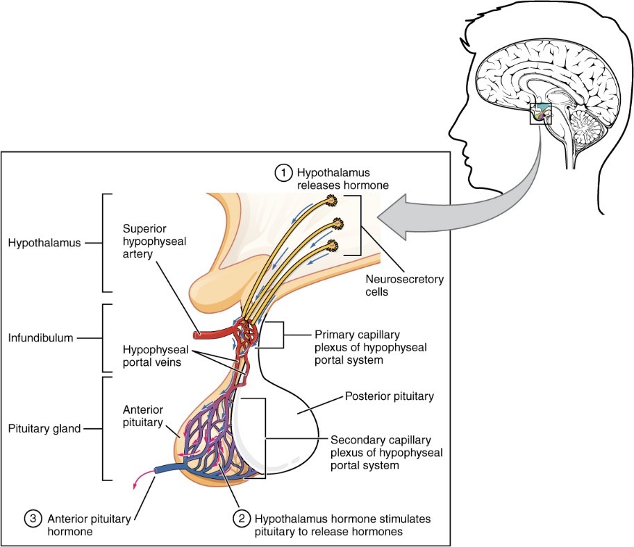
The Hypophysis (or Pituitary Gland) is an Endocrine Gland localised in the anterior part of the Brain where it is linked to the Hypothalamus. The Anterior lobe, called Adenohypophysis has many roles among others with 5 main hormones that it produces:

  • The stimulation of the Thyroiodin hormone’s secretion, essential to cell development, growth and differentiation (Thyrotropin (TSH))
  • Stress Response (Adrenocorticotropin (ACTH))
  • Milking, Reproductive and Behavioural functions (Gonadotrophins (FSH et LH) and Prolactin (PRL))
  • Cell Growth and Division (Growth Hormone (GH))


In partnership with Imagine Institute, we decided to model the Adenohypophysis because of the major role it plays in the Neuroendocrinal System and the organism’s metabolism. Additionally, its malfunction can induce many pathologies such as adenomas, pituitary dwarfism, gigantism, Cushing disease (obesity with diabetes, asthenia and hypertension) together with many disorders such as amenorrhea, sexual disorders, infertility, impotence, hypotension etc.

Only two models of pituitary organoids have been published during the past 10 years. The development of such an organoid represents a major challenge in the field of regenerative medicine, fundamental and pharmacological research.

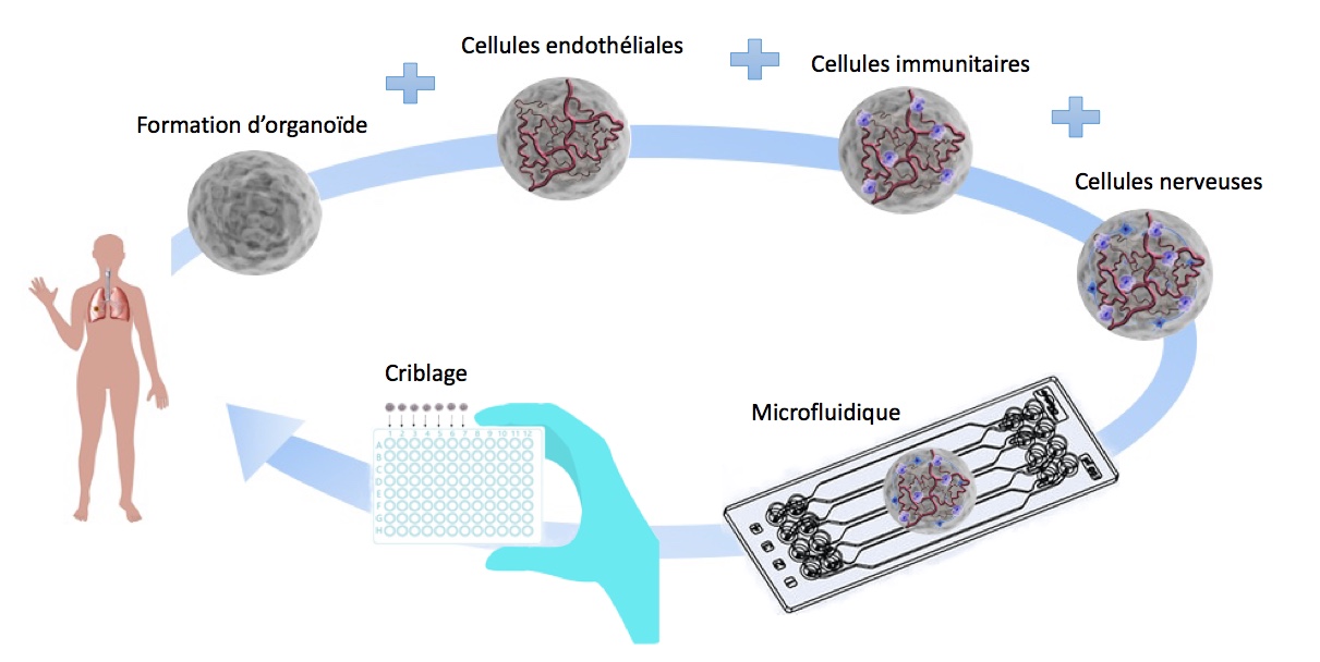
To create this Organoid, we will reproduce the Embryonic development of the Adenohypophysis by differentiating Human Induced Pluripotent Stem Cells (IPS) cultured in suspension into Oral Ectoderm then into Cranial placodes until the final pituitary cell subtypes. Our goal will be to reproduce these steps in an encapsulation microfluidic device to improve the reproductivity, reduce material costs and facilitate our experiments.

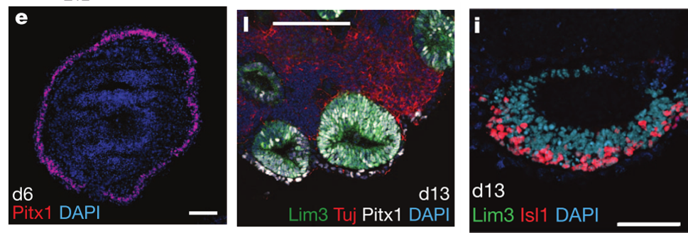
Figure 1. e. Neurectoderm formation (Day 6) l. Vesicles formation (Day 13) i. Rathke Pouch formation from the vesicles (Day 13) (Suga et al, 2011)


Retina Project

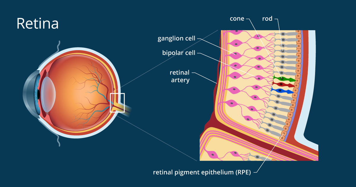
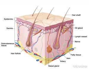
The retina is the innermost tissue and photosensitive epithelium of the eye. It is composed of two layers of different embryological origin, the nerve retina responsible for the absorption of the light signal and the pigmentary retina ensuring the conversion of the light into a bioelectric signal, transmitted mainly to the visual cortex by the optic nerve.

In partnership with Pasteur Institute, we have chosen to model the retina for its major medical issues in an aging society whose prevalence of retinal pathologies is gradually increasing. More than 170 million people are now affected by age-related macular degeneration (DMA) (Pennington & DeAngelis, 2016), a progressive deterioration of the area responsible for maximum visual acuity inducing a permanent spot in the centre of the field visual, frequently occurring in 65 years old people.

In recent years, several models of retinal organoids with different levels of complexity have been developed, with a significant structural and functional variability within and between models. They reveal some difficulties in terms of yield, functionality (especially on photoreceptors) including the presence of non-retinal structures.

Our model aims to improve the regularity, reproducibility and functionality of the retinal Organoid. Thus, to create this Organoid, we will reproduce the steps of Embryonic development of the retina by differentiating murine embryonic stem cells and induced human pluripotent cells (IPS). They will be cultured in a microfluidic system, to allow miniaturization and automation of cell culture procedures, enabling us to fulfil our objective of optimised reproducibility.

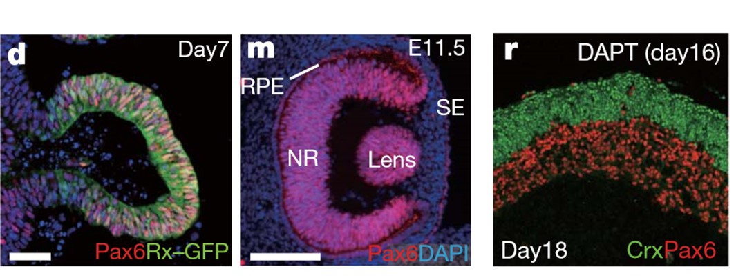
Figure 2 d.Formation of the vesicle (Day 7)   m.Formation of an optic-cup-like structure (Day 16),  r.Stratified neural retina tissues  (Day 11.5)  (Eiraku et al, 2011)


Sponsors

Members

Julie Jardon, Camille Brouillon, Guillaume Mondon, Nessim Richard